Home

délicat Intention Patate doppler varice casser OIE Pays

Jambes lourdes et varices | Clinique La Croix du Sud

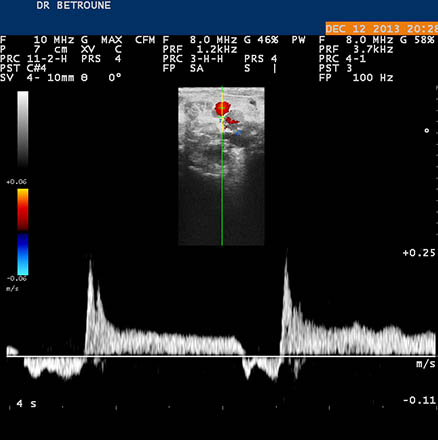
echod-doppler-varices - Dr Kahina Betroune

echodoppler-varice - Dr Kahina Betroune

Promo: Ecografia Doppler vasculară | FizioMed - Centru Medical Suceava

Varicose Veins of the Lower Extremity: Doppler US Evaluation Protocols,  Patterns, and Pitfalls | RadioGraphics
Varicose Veins of the Lower Extremity: Doppler US Evaluation Protocols, Patterns, and Pitfalls | RadioGraphics
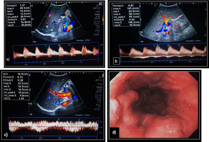
Figure 3 | Endoscopic Color Doppler Ultrasonography for Esophagogastric  Varices
Figure 3 | Endoscopic Color Doppler Ultrasonography for Esophagogastric Varices

Técnica y aplicaciones de la ecografía Doppler

Ecografia vascular | Fisi-on

Role of portal color Doppler ultrasonography as noninvasive predictive tool  for esophageal varices in cirrhotic patients | Egyptian Journal of  Radiology and Nuclear Medicine | Full Text
Role of portal color Doppler ultrasonography as noninvasive predictive tool for esophageal varices in cirrhotic patients | Egyptian Journal of Radiology and Nuclear Medicine | Full Text
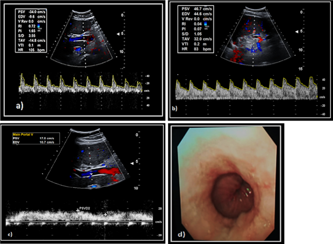
Varix | Eco Doppler Color

Écho Doppler Cartographie Des Varices - Centre MJ Médical - Marrakech

Phlébologue : quand et pourquoi consulter ?

Ecografie Doppler Vasculara Preventie Varice, program inclusiv in weekend

Role of portal color Doppler ultrasonography as noninvasive predictive tool  for esophageal varices in cirrhotic patients | Egyptian Journal of  Radiology and Nuclear Medicine | Full Text
Role of portal color Doppler ultrasonography as noninvasive predictive tool for esophageal varices in cirrhotic patients | Egyptian Journal of Radiology and Nuclear Medicine | Full Text

4-Modèle biomécanique avec une veine débordante. (a) Image Écho-Doppler...  | Download Scientific Diagram
4-Modèle biomécanique avec une veine débordante. (a) Image Écho-Doppler... | Download Scientific Diagram
3-Acquisition des images écho-doppler en position allongée (a) et... |  Download Scientific Diagram
3-Acquisition des images écho-doppler en position allongée (a) et... | Download Scientific Diagram

Diagnostics | Free Full-Text | Endoscopic Color Doppler Ultrasonographic  Evaluation of Gastric Varices Secondary to Left-Sided Portal Hypertension
Diagnostics | Free Full-Text | Endoscopic Color Doppler Ultrasonographic Evaluation of Gastric Varices Secondary to Left-Sided Portal Hypertension

Color doppler endoscopic ultrasound image of duodenal varices after... |  Download Scientific Diagram
Color doppler endoscopic ultrasound image of duodenal varices after... | Download Scientific Diagram

Examen d'imagerie médicale : Écho-doppler veineux des membres inférieurs |  ameli.fr | Assuré
Examen d'imagerie médicale : Écho-doppler veineux des membres inférieurs | ameli.fr | Assuré

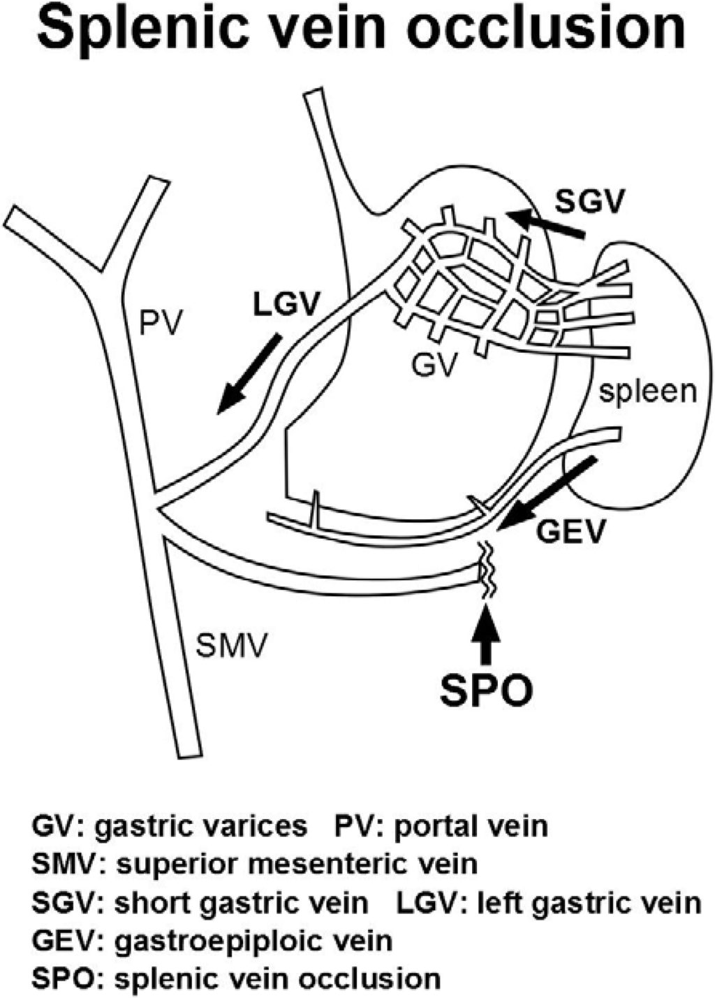
Endoscopic color Doppler ultrasonography of gastric variceal flow... |  Download Scientific Diagram
Endoscopic color Doppler ultrasonography of gastric variceal flow... | Download Scientific Diagram
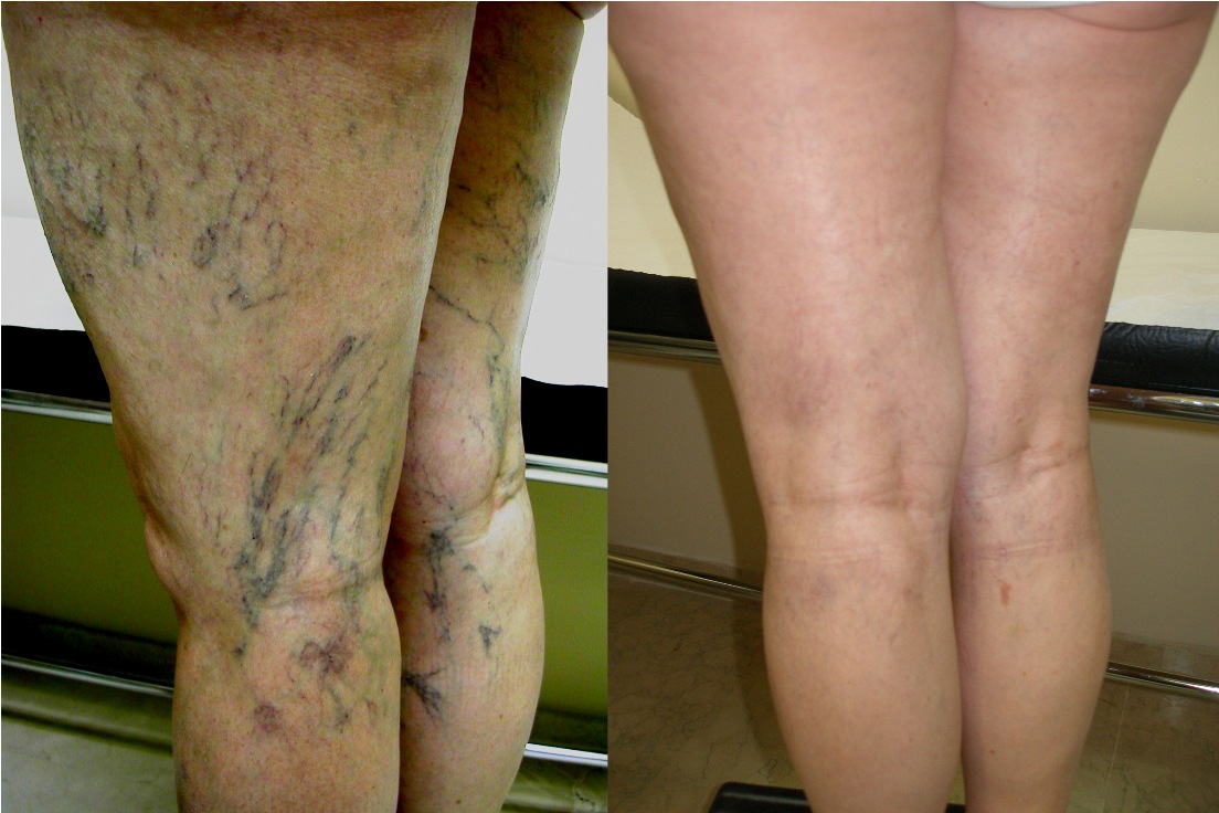
Loosli & Coll. : Angiologues et phlébologues à Marseille

Ecografía doppler es clave para conocer las causas de las várices - Clínica  Las Condes
Ecografía doppler es clave para conocer las causas de las várices - Clínica Las Condes

Specific criteria of the transcutaneous Doppler ultrasound in unusual  causes of lower limb varicose veins - Servier - PhlebolymphologyServier –  Phlebolymphology
Specific criteria of the transcutaneous Doppler ultrasound in unusual causes of lower limb varicose veins - Servier - PhlebolymphologyServier – Phlebolymphology